The Brain in Hypnosis
The Brain is Not the Mind
When considering brain waves and neuroimaging of hypnosis, first keep in perspective that changes in electrical activity in the brain do little to explain the subjective experience and intentionality of consciousness.
There are two basic ways of looking at the relationship between the brain and the mind:
The physicalist viewpoint argues that electrical impulses in the brain create the subjective experience we call the “mind,” and that consciousness is a product of the continuous activity of the brain.
Others believe that mind is a property of nature (like electrical charge, spin, or mass) which exists independent of the brain, and that the mind interacts with the brain as a device that it uses and directs. Dr. Wilder Penfield (1891-1976), the renowned father of modern neurosurgery, reached the conclusion that the mind has a reality of its own, far more than being a product of the brain. In Mystery of the Mind: A Critical Study of Consciousness and the Human Brain he wrote:
“The mind seems to act independently of the brain in the same sense that a programmer acts independently of his computer, however much he may depend upon the action of that computer for certain purposes…
“To expect the highest brain-mechanism or any set of reflexes, however complicated, to carry out what the mind does, and thus perform all the functions of the mind, is quite absurd..
“It seems to me certain that it will always be quite impossible to explain the mind on the basis of neuronal action within the brain…
Brain Wave Patterns
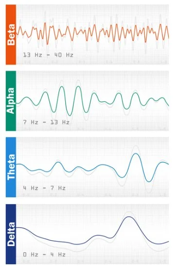
Humans have four basic states of awareness corresponding to four identifiable brain-wave patterns:
Beta state (alertness, active thinking, and concentration)
Alpha state (relaxed alertness and light hypnosis)
Theta state (day dreaming, deep hypnosis, drowsiness, and light sleep)
Delta state (deep sleep).
The brain passes through each of these four states as the electrical activity decreases on the way to sleep (theta and delta states) and increases on the way to wakefulness (beta state). In other words, we necessarily pass through the hypnotic states every time we go into and awaken from sleep.
We spend most of our waking hours in the beta state of alert awareness. In the beta state the mind analyzes, evaluates, judges, and makes decisions. This is the state in which we attempt to overcome problems with “will power,” often unsuccessfully.
In hypnosis the brain enters the alpha (light hypnosis) and theta (deep hypnosis) states, and one is highly focused on hypnotic suggestions and imagery while suspending the ordinary thinking processes of the beta state. In the alpha and theta states, hypnotic suggestions are integrated into the mind more easily, and memories become more accessible.
There are many techniques for inducing hypnosis, and most of them bring about the alpha and theta hypnotic states with the same basic method that you use to put yourself to sleep at night: you close your eyes, control the sound, and lie still. When you limit sensory stimulation in these ways, your neural activity slows down, taking you from the waking state (beta) into the states of hypnosis (alpha and theta). In these states the brain’s centers of awareness and imagining shift from cortex (where conscious, analytical thinking takes place) to the sub-cortical structures involved in unconscious and emotional processes, the stress response, and long-term memory. In simple terms, by limiting sensory input the brain slows down, and the unconscious mind is made accessible.
Neuroimaging of Hypnosis
Whether hypnosis is an altered state of consciousness has been debated for nearly 200 years. Today, brain imaging studies seem to confirm that hypnosis is an altered state. Numerous studies using electroencephalography (EEG), positron emission tomography (PET), and functional magnetic resonance imaging (fMRI) show distinct changes in the brain during hypnosis and in response to suggestion.
Brain imaging studies observing the general effect of the hypnotic state (as opposed to the effects of specific suggestions) have found that hypnosis causes observable changes in the brain areas and systems involved in:
Consciousness and sense of self. [1]
Attentional absorption and spontaneous conceptual thought. [2]
Concentration, attentional control and executive function (reasoning, problem solving, planning, self-control, and cognitive flexibility). [3]
Higher cortical functions. [4]
Awareness and control of internal bodily processes and emotions. [11]
Emotional evaluation and worrying. [11]
Studies observing the effects of hypnotic suggestions have most frequently centered on the neurophysiological response to suggestions for pain, motor function and limb paralysis, mental imagery, and memory. These studies demonstrate not only that changes occur in brain physiology, but also that these changes explain the phenomena and behaviors observed in hypnosis. Hypnotic suggestion has been shown to affect the brain areas and systems involved in:
Consciousness, sleep, and alertness. [5]
Motor control. [6]
Autonomic functions, emotion, motivation, impulse control, reward anticipation, and decision-making. [7]
Color perception. [8]
Reasoning and decision making. [9]
Imagery, self-awareness, and motor control. [10]
Neural activity in hypnosis
Hypnosis causes changes in brain activity and connectivity consistent with decreased self-consciousness, increased control of internal sensations and emotion, and less worry.
A study by researchers at Stanford University in July 2016 considered what takes place in the brain in general during hypnosis. Functional magnetic resonance imaging (fMRI) was used to observe brain activity in 57 subjects in hypnosis. Changes were observed in three specific areas of the brain:[11]
1. Reduced connectivity between the dorsolateral prefrontal cortex (part of the executive control network involved in planning and decision making) and the posterior cingulate cortex (the part of the default mode network that seems to be involved self-related thinking and one's sense of self). This may be responsible for the dissociation that can occur in hypnosis, enabling one to place certain events, thoughts, and sensations outside of the self and thus permit a shift in the cognitive set (i.e. belief system).
The inverse functional connectivity between regions involved in planning of actions and those involved in self-awareness may also account for the immediacy of action that takes place during hypnosis. In hypnosis, as in states of deep absorption in a task or performance, one acts spontaneously, without reflecting upon actions.
2. Increased connectivity between the dorsolateral prefrontal cortex (part of the executive control network involved in planning and decision making) and the insula (involved in sensing and regulating autonomic internal bodily processes). This may be why in hypnosis one has increased control over autonomic process (e.g.: pain perception, blood flow, temperature, dilation of the pupils).
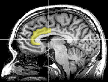
3. Reduced activity in the dorsal anterior cingulate cortex (dACC). This is part of the salience network, which helps us to notice things that stand out to our attention (e.g.: when you see something out of place, hear a strange noise, or feel an odd sensation). The dACC is involved in the emotional evaluation of errors and worrying, and is active during effortful performance. Reduced activity in the dACC may explain the high level of focus that characterizes hypnosis, and why actions and performance take place effortlessly and with less worry in states of hypnosis and absorption (i.e. "flow").
Reduced activity in parts of the brain's default mode network (DMN) increases attentional absorption.
The DMN is a network of interacting brain regions that is active when a person is not involved in a task, and when thinking, remembering, and daydreaming. The DMN is not active when a person is involved in a goal-oriented task or has their attention completely absorbed by something.
Hypnosis reduces the activity of the DMN.[2] Reduced activation of the DMN is also observed in long-term practitioners of meditation. The fact that the brain under hypnosis shows reduced DMN activity supports the definition of hypnosis as a state of attentional absorption, rather than a state where one loses consciousness. Hypnosis shows neural responses similar to spontaneous conceptual thought. Perhaps this is why hypnosis can spark creativity and insight.
Default Mode Network (DMN).
Increased activity in the prefrontal attentional systems increases executive function.
The frontal areas of the brain govern a person’s ability to concentrate, known as attentional control. Attentional control is closely related to executive functions such as reasoning, problem solving, planning, self-control, and cognitive flexibility (the ability to think about multiple concepts at the same time). Hypnosis increases the activity of the systems involved in these functions, and which are impaired in addiction and ADHD.[2]
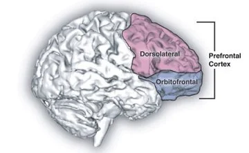
Prefrontal cortex.
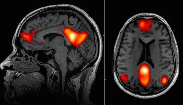
Hypnosis modulates activity in the brain areas involved in the regulation of consciousness.
PET scans of cerebral blood flow in hypnosis show the involvement of the anterior cingulate cortex (ACC), the thalamus, and the ponto-mesencephalic brainstem. Hypnosis increases blood flow in the occipital region, which is consistent with the theory that hypnosis decreases cortical arousal (i.e., cortical inhibition theory). Increases in mental absorption during hypnosis were associated with increased blood flow in the brain's attentional system.[4]
Anterior cingulate cortex (ACC).
Sources:
[1] Rainville, P., & Price, D. D. (2003). Hypnosis Phenomenology and the Neurobiology of Consciousness. International Journal of Clinical and Experimental Hypnosis, 51(2), 105-129.
[2] Deeley, Q., Oakley, D. A., Toone, B., Giampietro, V., Brammer, M. J., Williams, S. C., & Halligan, P. W. (2012). Modulating the Default Mode Network Using Hypnosis. International Journal of Clinical and Experimental Hypnosis, 60(2), 206-228.
[3] Ibid.
[4] Rainville, P., Hofbauer, R. K., Bushnell, M. C., Duncan, G. H., & Price, D. D. (2002). Hypnosis Modulates Activity in Brain Structures Involved in the Regulation of Consciousness. Journal of Cognitive Neuroscience, 14(6), 887-901.
[5] Müller, K., Bacht, K., Prochnow, D., Schramm, S., & Seitz, R. J. (2013). Activation of thalamus in motor imagery results from gating by hypnosis. NeuroImage, 66, 361-367.
[6] Ibid.
[7] Vanhaudenhuyse, A., Boly, M., Balteau, E., Schnakers, C., Moonen, G., Luxen, A., . . . Faymonville, M. (2009). Pain and non-pain processing during hypnosis: A thulium-YAG event-related fMRI study. NeuroImage, 47(3), 1047-1054.
[8] Kosslyn, S. M., Thompson, W. L., Costantini-Ferrando, M. F., Alpert, N. M., & Spiegel, D. (2000). Hypnotic Visual Illusion Alters Color Processing in the Brain. American Journal of Psychiatry AJP, 157(8), 1279-1284.
[9] Raij, T. T., Numminen, J., Närvänen, S., Hiltunen, J., & Hari, R. (2009). Strength of prefrontal activation predicts intensity of suggestion-induced pain. Human Brain Mapping Hum. Brain Mapp., 30(9), 2890-2897.
[10] Cojan, Y., Waber, L., Schwartz, S., Rossier, L., Forster, A., & Vuilleumier, P. (2009). The Brain under Self-Control: Modulation of Inhibitory and Monitoring Cortical Networks during Hypnotic Paralysis. Neuron, 62(6), 862-875.
[11] Jiang, H., White, M. P., Greicius, M. D., Waelde, L. C., & Spiegel, D. (2016). Brain Activity and Functional Connectivity Associated with Hypnosis. Cerebral Cortex.